Plant Cell Microscope Slide - Typical Plant Cell 100x Dissection Connection - The microscope slides are first washed.. Tissue from an onion is a good first exercise in using the microscope and viewing plant cells. Light and electron microscopes allow us to see inside cells. A cell wall for strength a large vacuole for storage chloroplasts for photosynthesis. This appears at the light microscope level as a duplication of chromosomes. Preparing animal and plant cell slides (examples.
See more ideas about microscopic photography, plant cell, microscopic. Science supplies science kits microscope slides plant cell stem cells homeschool delicate activities. Tissue from an onion is a good first exercise in using the microscope and viewing plant cells. Microscope slide cover slip onion. A list of stains used on microscope slides, and what the differences between them are.
3 576 Microscope Slide Stock Videos And Royalty Free Footage Istock from media.istockphoto.com A simple and inexpensive micrometer slide may be made by mounting a 200 mesh electron microscope grid on a microscope slide under a coverslip. Science supplies science kits microscope slides plant cell stem cells homeschool delicate activities. Unroll the specimen so that it is flat, and place it in the middle of a clean slide. Just place your prepared slide of plant between light and slide stand and focus on 40x or 100x you can easily see plant cells under microscope. To examine plant cells under a microscope and find and identify different cell parts. Please send one lab person to retrieve • 2. Before cell division, the entire genome is copied. Preparing animal and plant cell slides (examples.
With careful handling, living plant cells can be used very effectively on the confocal microscope.
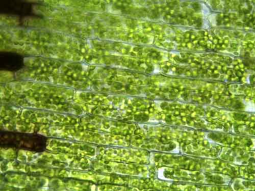
A plant's perspective on cell polarity, cell fate transitions and intercellular communication. And she coughs, which is kind of gross. To examine plant cells under a microscope and find and identify different cell parts. Plant cells with green chloroplasts under microscope. Plant cell microscope slide labeledshow all. Green chlorophyll, chloroplasts in plant eukaryotic cell structures, magnification in microscope. The premier plant biology microscope slides choices on alibaba.com add incredible accuracy in lab analysis. Cut a thin section of stem or leaf which you want to observe. Ever since the first microscope was used, biologists have been interested in studying the cellular. It prevents the slide from drying out when it's being. Place the slide under the. Animal and plant cells undergo a precise type of division called mitosis. Onion cellsgreen plant cells animal cells.
Green chlorophyll, chloroplasts in plant eukaryotic cell structures, magnification in microscope. This is one of the tenets of the cell theory, a basic theory of biology. A simple and inexpensive micrometer slide may be made by mounting a 200 mesh electron microscope grid on a microscope slide under a coverslip. The microscope slides are first washed. Animal cells introduction background information:
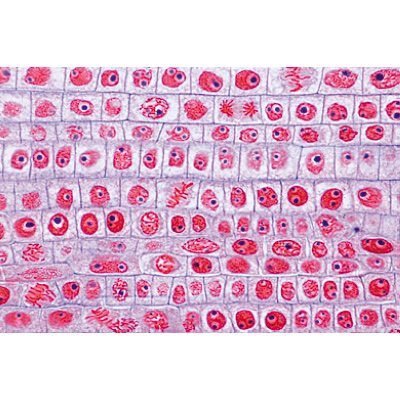
General Biology Microscopic Specimen Images Photographs from www.scienceprofonline.com A visual tour of the building block of life. The thin membrane from between the layers of a raw onion provides a good material for viewing plant cells. All living things are composed of cells. Place the slide under the. Slide preparation onion cell slides are prepared with iodine instead of water. iodine stains the membrane and nucleus to make it visible. green plant prepared with water. animal (cheek) cell slides prepared with blue. Animal and plant cells undergo a precise type of division called mitosis. Plant cell microscope slide labeledshow all. A plant's perspective on cell polarity, cell fate transitions and intercellular communication.
Microscope slide of onion (allium) bulb epidermis, used to study the general structure of plant cells. Light and electron microscopes allow us to see inside cells. Outside skin of an onion. A brief treatment with cell wall degrading enzymes is often used to. With careful handling, living plant cells can be used very effectively on the confocal microscope. Please send one lab person to retrieve • 2. My students used to love microscope lab days. 1 box, 50 pcs/box colour:white material:plastic + glass size:76.2*25.4*1mm package contents: It prevents the slide from drying out when it's being. Animal cells introduction background information: The thin membrane from between the layers of a raw onion provides a good material for viewing plant cells. In plant cells, a cell plate is formed in the middle of the spindle and later extends across the whole cell. The images showcase the building blocks of the organisms around us.
A small square or circle of thin glass called a coverslip is placed over the specimen. Place the slide under the. Preparing animal and plant cell slides (examples. Unroll the specimen so that it is flat, and place it in the middle of a clean slide. Preparing onion cell slides is a useful way to observe simple plant cells under the light microscope.
Plant Cell English Slides 1003982 W13053 Lieder 5100 En English 3b Scientific from www.3bscientific.com 50 pcs prepared slides quantity: Plant cells have three features not present in animal cells: From our beginnings in 1927, carolina biological supply company has grown to become a leading supplier of science teaching materials for all levels of education. Photo about microscopic view of a plant stem cross cut section under the scientific microscope. The microscopy slides are part of a recently released book called the cell: Animal and plant cells undergo a precise type of division called mitosis. An onion is made of layers, each separated by a thin skin or membrane. Science supplies science kits microscope slides plant cell stem cells homeschool delicate activities.
An onion is made of layers, each separated by a thin skin or membrane.
1 box, 50 pcs/box colour:white material:plastic + glass size:76.2*25.4*1mm package contents: A small square or circle of thin glass called a coverslip is placed over the specimen. Slide preparation onion cell slides are prepared with iodine instead of water. iodine stains the membrane and nucleus to make it visible. green plant prepared with water. animal (cheek) cell slides prepared with blue. A brief treatment with cell wall degrading enzymes is often used to. Microscope slide cover slip onion. The microscopy slides are part of a recently released book called the cell: A view through the microscope. Ever since the first microscope was used, biologists have been interested in studying the cellular. Găsește imagini de stoc cu tema plant cell microscope slide real shot, în format hd, și alte milioane de fotografii, ilustrații și vectori de stoc fără drepturi de autor din colecția shutterstock. See more ideas about microscopic, plant cell, microscopic photography. Stains acidic cell parts (like nucleus) blue. The thin membrane from between the layers of a raw onion provides a good material for viewing plant cells. Place it on a slide and put a small amount of colouring agent.
My students used to love microscope lab days plant cell microscope. A view through the microscope.
Share :
Post a Comment
for "Plant Cell Microscope Slide - Typical Plant Cell 100x Dissection Connection - The microscope slides are first washed."


Post a Comment for "Plant Cell Microscope Slide - Typical Plant Cell 100x Dissection Connection - The microscope slides are first washed."